It’s True – Metals in a Tattoo – Systemic Contact Dermatitis

Yes, indeed metals can be implanted in a tattoo… and systemically activated reactions can occur in those tattoos related to those metals…

“We recommend assessment of permanent tattoos for inflammation in all patients undergoing patch testing, for additional diagnostic correlation.”  [ 2008 Sep-Oct;19(5):E33-4]


Article 1 (came out yesterday) :de Cuyper C1Lodewick E2Schreiver I3Hesse B4Seim C5,6Castillo-Michel H4Laux P3Luch A3.   2017 Aug 9. doi: 10.1111/cod.12862. [Epub ahead of print]  Are metals involved in tattoo-related hypersensitivity reactions? A case report.

“BACKGROUND:  Allergic reactions to tattoos are not uncommon. However, identification of the culprit allergen(s) remains challenging.

OBJECTIVES: We present a patient with papulo-nodular infiltration of 20-year-old tattoos associated with systemic symptoms that disappeared within a week after surgical removal of metal osteosynthesis implants from his spine. We aimed to explore the causal relationship between the metal implants and the patient’s clinical presentation.

METHODS: Metal implants and a skin biopsy of a reactive tattoo were analysed for elemental contents by inductively coupled plasma mass spectrometry and synchrotron-based X-ray fluorescence (XRF) spectroscopy.

RESULTS: Nickel (Ni) and chromium (Cr) as well as high levels of titanium (Ti) and aluminium were detected in both the skin biopsy and the implants. XRF analyses identified Cr(III), with Cr(VI) being absent. Patch testing gave negative results for Ni and Cr. However, patch tests with an extract of the implants and metallic Ti on the tattooed skin evoked flare-up of the symptoms.

CONCLUSION: The patient’s hypersensitivity reaction and its spontaneous remission after removal of the implants indicate that Ti, possibly along with some of the other metals detected, could have played a major role in this particular case of tattoo-related allergy.”


Article 2: Cobalt tattoo reaction:

2017 Jun 1;15(3):221-222. eCollection 2017.  Chemical Tattoo Treatment Leading to Systemic Cobalt Hypersensitivity.  Zajdel NJ1, Smith WA2, Taintor AR3, Jacob SE4, Olasz EB5.

“An otherwise healthy 36-year-old Caucasian woman, without prior history of atopic dermatitis or eczema, presented to an outside dermatologist with a generalized, severely pruritic eruption involving the entire body except the face. One month previously, she had used a 50% trichloroacetic acid tattoo removal solution on a blue-colored tattoo on the medial aspect of the left ankle. The patient’s eruption persisted for 7 months, and after several attempts to slowly taper her prednisone dose, she presented to our institution. On physical examination, there was a 3-cm erythematous, lichenified plaque surrounding the tattoo (Figure). On the trunk and upper regions of the arms, there were scattered, 1- to 2-cm, nummular patches and plaques. Biopsy of a truncal lesion revealed spongiotic pustules with a mixed dermal infiltrate and scattered eosinophils, consistent with subacute spongiotic dermatitis.”


Article 3:  Systemic Dermatitis following surgery — presenting as tattoo reaction

2017 Jul 19;3(4):348-350. doi: 10.1016/j.jdcr.2017.05.003. eCollection 2017 Jul.  Systemic contact dermatitis to a surgical implant presenting as red decorative tattoo reaction.
“The patient reported that within 2 weeks of surgery, the red-containing areas of her tattoos, which were previously flat and uninflamed, became raised and pruritic.”…
Read this article
Article 4: SCD to chromate in a tattoo triggered by patch testing
2008 Sep-Oct;19(5):E33-4.Inflammation in green (chromium) tattoos during patch testing.  Jacob SE1, Castanedo-Tardan MP, Blyumin ML.

“We report three patients with permanent tattoos and chronic dermatitis. During patch testing, the patients’ dermatitis worsened, and the previously quiescent green-colored portions of the tattoos became inflamed. All three patients were patch-tested and had positive reactions to potassium dichromate 0.25% in petrolatum. Avoidance led to the resolution of both the dermatitis and the tattooinflammation. We recommend assessment of permanent tattoos for inflammation in all patients undergoing patch testing, for additional diagnostic correlation.”

Article 5: 1962
1962 Aug-Sep;74:288-94.Green tattoo reactions associated with cement dermatitis.
And … one of those reads that just makes you think… Article 6:
2004 Aug 21-27;364(9435):730.  A red tattoo and a swordfish supper.
Read more here
“Tsuruta et al. report- ed a case of a 40-year-old Japanese man with a red tattoo who developed a whole-body rash after eating 250 g of raw swordfish and alfonsino.”
Researchers are investigating metal allergic dermatitis and the role of piercing in nickel allergy.  Please pass along this survey.

airborne allergic contact dermatitis- isothiazolinones is not rare

2017 Apr 27. doi: 10.1111/cod.12795. [Epub ahead of print]

Airborne allergic contact dermatitis caused by isothiazolinones in water-based paints: a retrospective study of 44 cases.

Abstract

BACKGROUND:

Airborne allergic contact dermatitis caused by paints containing isothiazolinones has been recognized as a health hazard.

OBJECTIVES:

To collect epidemiological, clinical and patch test data on airborne allergic contact dermatitis caused by isothiazolinone-containing paints in France and Belgium.

METHODS:

A descriptive, retrospective study was initiated by the Dermatology and Allergy Group of the French Society of Dermatology, including methylchloroisothiazolinone (MCI)/methylisothiazolinone (MI)- and/or MI-sensitized patients who developed airborne allergic contact dermatitis following exposure to isothiazolinone-containing paint.

RESULTS:

Forty-four cases were identified, with mostly non-occupational exposure (79.5%). Of the patients, 22.5% of also had mucosal symptoms. In several cases, the dermatitis required systemic corticosteroids (27.3%), hospitalization (9.1%), and/or sick leave (20.5%). A median delay of 5.5 weeks was necessary to enable patients to enter a freshly painted room without a flare-up of their dermatitis. Approximately one-fifth of the patients knew that they were allergic to MI and/or MCI/MI before the exposure to paints occurred.

CONCLUSION:

Our series confirms that airborne allergic contact dermatitis caused by paints containing isothiazolinones is not rare, and may be severe and long-lasting. Better regulation of isothiazolinone concentrations in paints, and their adequate labelling, is urgently needed.

https://www.ncbi.nlm.nih.gov/pubmed/28449346

Free Article on Topical Steroid addiction – withdrawal

Free article on topical steroid addiction – “Cortisol production by keratinocytes [skin cells] might work to regulate or moderate the friction between the outer environment and inner immune system by suppressing excessive inflammation or immune reaction. However, prolonged or excessive use of TCS induces skin atrophy which can make barrier function weak. Moreover, the decreased self-production of cortisol by the keratinocytes can cause hypersensitivity. The author considers it is one of the mechanisms of TSA or rebound phenomenon after TSW.”

Fukaya M1. .Histological and Immunohistological Findings Using Anti-Cortisol Antibody in Atopic Dermatitis with Topical Steroid Addiction.   2016 Mar;6(1):39-46. doi: 10.1007/s13555-016-0096-7. Epub 2016 Feb 2.  Dermatol Ther (Heidelb)

“Abstract

INTRODUCTION:

Though topical steroid addiction (TSA) in patients with atopic dermatitis (AD) has been recently discussed as a clinical problem, there are very few studies about its mechanism. The purpose of this study was to elucidate histological and immunohistological characteristics of TSA using anti-cortisol antibody.

METHODS:

Skin biopsy specimen from eight patients with AD was stained by anti-cortisol antibody (Biorbyt, orb79379). Subjects consisted of a child patient with a short history of topical corticosteroids (TCS) application, an adult patient with a long history of TCS application, and six adult patients who have experienced topical steroid withdrawal (TSW) and the rebound phenomenon.

RESULTS:

The staining in the epidermis by anti-cortisol antibody presented patchy defects in the child patient, the patient with a long history of TCS application, and two patients at the rebound period. Parakeratosis with poor formation of corneal layer was obvious in the child patient, the patient with a long history of TCS application, two patients recovered from TSA, and two patients at the rebound period.

CONCLUSION:

Prolonged application of TCS might suppress the cortisol production of keratinocytes which is poorly developed at the early ages before childhood and completed naturally as to growth. Rebound phenomenon after TSW can occur due to the relative insufficiency of cortisol in the epidermis and the immature corneal layer formation.”

https://www.ncbi.nlm.nih.gov/pmc/articles/PMC4799038/

Contact Dermatitis Awareness Ribbon

Announcing the Contact Dermatitis Awareness Ribbon:

On March 19, 2016 a Montessori Teacher and a Customer Service Representative, two mothers with children suffering with allergic contact dermatitis, joined together to start a patient-centered outreach group on Facebook called “Eczema, Contact Dermatitis and Patch Testing Alliance”. Currently, this 1,925 member focus group is providing educational resources to sufferers of allergic contact dermatitis worldwide.

As the eve of the anniversary of the group approached the lead administrator (Misha Bertolino, MA) raised the question, Why is there not a contact dermatitis awareness ribbon?

**Contact dermatitis costs a reported $1,529 million/year in medical costs!

**Contact dermatitis is the 8th most costly skin disease!

**Contact dermatitis is preventable!


The Contact Dermatitis Awareness Ribbon is indeed very much needed!

In a collaborative effort, the Eczema, Contact Dermatitis and Patch Test alliance along with artist Janna Vassantachart, MD, logistician Chandler Rundle, BS, practicing contact dermatitis specialists, and global advocates – the orchid (eczema) and teal (allergy) contact dermatitis awareness ribbon has become a reality.

This symbol can be worn to show support and solidarity for the millions of people who suffer from this disease.  In alignment with these symbols, our mission at the Dermatitis Academy is to educate the public, the medical providers, the manufacturers and the legislators on ACD, while cultivating a community of support for those impacted by this disease.

With early diagnosis, education, and intervention, we HOPE for a future where allergic contact dermatitis can be controlled by remission or prevention.

Please visit the Dermatitis Academy to learn more about allergic contact dermatitis, allergens, and patch testing and to download the Contact Dermatitis Awareness Ribbon.

Please share!

Nickel sensitivity and atopy

Early insight into the contributory role genetic factors play in the development of contact dermatitis

Synopsis of Nickel Sensitivity and Atopy (1964)

Original article:

A. Caron. (1964). Nickel Sensitivity and Atopy. British Journal of Dermatology, vol. 76: p. 384 – 387.

Reviewed by Brittanya Limone, BS, MA. MSIII, Loma Linda University.

  • “Atopy” is a genetic predisposition toward developing one or more of the following conditions: asthma, hay fever, urticaria, infantile eczema, or atopic dermatitis.
  • Determining the incidence of atopy has proven difficult because of reliance on clinical judgment and variable utilization of set parameters in making the diagnosis. For instance, the presentation of nasal congestion and recurrent sneezing episodes may be diagnosed as either hay fever, allergic rhinitis, or vasomotor rhinitis.
    • Several authors c.1964 estimated the prevalence of atopy in the United States’ general population being between 10-20%.
  • In the Caron case series, 37 patients with an established diagnosis of nickel contact dermatitis underwent further evaluation regarding their personal and family history of atopy, the results included the following:
    • Twenty-one (54%) had no history of atopy
    • Seven (19%) only had a family history.
    • Four (11%) had both a personal and family history
    • Five (14%) only had a personal history
  • The results from Caron’s case series suggested that the incidence of atopy amongst patients with nickel allergic contact sensitivity was no greater than its occurrence in the general population.
    • The frequency of atopy in 19% of family members was noted to be higher than expected for the general population but was attributed to the contributory role genetic factors play in the development of contact dermatitis.
  • This study concluded that nickel sensitivity occurs independently of atopy.

https://www.ncbi.nlm.nih.gov/pubmed/14201189

Nickel Earlobe Dermatitis – persistent sensitivity!

Nickel Earlobe Dermatitis

Original article:

Thomas L. Watt and Robert R. Baumann. (1968). Nickel Earlobe Dermatitis. Archives of Dermatology, vol. 98: p. 155-158.

Reviewed by Brittanya Limone, BS, MA. MSIII, Loma Linda University.

  • Although nickel is not a high potency sensitizer, increased frequency of nickel exposure, especially amongst young women, make it a common cause of allergic contact dermatitis.
  • The development of nickel sensitization can result in persistent sensitivity despite avoidance of items containing the metal. In addition, some patients may later have negative nickel patch testing but continue to have a dermatitis that stemmed from the nickel allergy.
  • Following repeated exposures, early nickel intolerances can eventually lead to metal rejection across the skin’s surface. This may lead to secondary site dermatitis reactions or a generalized dermatitis in some patients.
  • Watt and Baumann conducted a year-long observational study in 17 young women after they developed a draining ear lobe dermatitis 2-4 weeks after having ear lobe piercings with implanted earrings.
    • All of the women developed a positive eczematous reaction to nickel patch testing. Eleven had a personal history of atopy and three carried a family history.
    • Previously, the patients had tolerated nickel exposures to costume jewelry and clothing fasteners without trouble. The nickel allergy was only apparent following the earlobe piercing.
    • “In our experience, nickel sensitization following earlobe piercings is commonly mistaken for chronic infection, even though a similar dermatitis of the nonpierced earlobe is considered to be a cardinal sign of nickel allergy”.
  • The study found that despite the common perception that nickel dermatitis occurs more frequently after wearing “inexpensive” jewelry, nickel dermatitis was just as readily observed in these women following exposure to 14-karat gold and sterling silver earrings as with the inexpensively plated jewelry.
  • The study concluded that nickel earlobe dermatitis was highly associated with a personal history of atopy. Therefore, determination of a prior history of nickel allergy and atopy must be conducted prior to earlobe piercings.
    • Of note, careful screening for “latent atopics” must also be performed as they are equally susceptible to nickel earlobe dermatitis. These individuals may never have an obvious atopic disease but have a positive family history, positive skin testing, physical signs suggesting atopy, and bear atopic offspring.
  • According to Watt and Baumann, a history of nickel allergy is an “absolute contraindication” to earlobe piercings. On the other hand, personal history of atopic disease only requires warning patients about the possibility of nickel sensitization.

https://www.ncbi.nlm.nih.gov/pubmed/5667228

New data regarding Wet Wipe Allergens from the North American Contact Dermatitis Group!

New data regarding Wet Wipe Allergens from the North American Contact Dermatitis Group!

There have been several case reports of wipe-associated contact dermatitis, however, the aim of this study was to determine the prevalence of wipe-associated contact dermatitis in a larger population This study looked at 9037 patients patch tested from 2011-2014 to determine the prevalence of wet wipes as a source of contact allergy.

What did they find?

79(0.9%) had a positive patch test to an allergen associated with a wet-wipe source. Anal/genital dermatitis was 15 times more likely in those with a wet-wipe allergy!

What were the most associated allergens

Preservatives and fragrance:

  1. Methylisothiazolinone (59.0%)
  2. Methychloroisothiazolinone (MCI)/MI (35.6%)
  3. Bronopol (2-bromo-2-nitropropane-1,3-diol) (27.4%)
  4. Iodopropynyl butylcarbamate (12.3%)
  5. Fragrance (combined) represented (12.3%)

What was their conclusion?

Although uncommon (0.9%), Wet wipes are an important source of contact allergy to consider with anal/genital dermatitis. Preservatives such as Isothiazolinones are an especially important source to consider!


Warshaw EM, Aschenbeck KA, Zug KA, Belsito DV, Zirwas MJ, Fowler JF Jr, Taylor

JS, Sasseville D, Fransway AF, DeLeo VA, Marks JG Jr, Pratt MD, Maibach HI,

Mathias CG, DeKoven JG. Wet Wipe Allergens: Retrospective Analysis From the North

American Contact Dermatitis Group 2011-2014. Dermatitis. 2017

Jan/Feb;28(1):64-69

Nickel allergy and wheat sensitivity – Free access article

Contact Dermatitis Due to Nickel Allergy in Patients Suffering from Non-Celiac Wheat Sensitivity 

Original article:

Alberto D’ Alcamo, Pasquale Mansueto, Maurizio Soresi, Rosario Iacobucci, Francesco La Blasca, Girolamo Geraci, Francesca Cavataio, Francesca Fayer, Andrea Arini, Laura Di Stefano, Giuseppe Iacono, Liana Bosco & Antonio Carrocio. Contact Dermatitis Due to Nickel Allergy in Patients Suffering from Non-Celiac Wheat Sensitivity.Nutrients, 2017; Vol. 9(2):103  

Reviewed by Sue Min S. Kwon, BS, MSI and Annelise Rasmussen BS, MSII, Loma Linda University.

  • As gluten allergy becomes more prevalent and widely-knownin society, patients with cutaneous or gastrointestinal symptoms following wheat ingestion may self-report gluten/wheat allergies, though they do not in fact have celiac disease. Almaco et al. suggested the term “non-celiac wheat sensitivity” (NCWS) to describe patients presenting with these symptoms, rather than non-celiac gluten sensitivity (NCGS), as it is not known which component of wheat causes the symptoms.
  • Non-celiac wheat sensitivity (NCWS) is a relatively new clinical finding associated with gluten-related diseases. Wheat contains nickel, a known contact allergen, which may produce systemic nickel allergy syndrome (SNAS) symptoms. Nickel is the most frequent cause of contact allergy in tested populations.
  • NCWS can mimic irritable bowel syndrome (IBS). 
  • Almacoet al. conducted a double-blind placebo-controlled (DBPC) experiment in order to evaluate the frequency of contact dermatitis due to nickel allergy.

o   NCWS patients suffering from nickel allergy were compared with a control group of NCWS patients who did not report nickel allergy.

o   NCWS patients with nickel allergy had a significantly higher percentage of atopic disease manifestations than those with irritable bowel syndrome (IBS) and NCWS patients without nickel allergy.

  • Nickel allergy (diagnosed by a confirmatory epicutaneouspatch test) may manifest with both cutaneous and gastrointestinal symptoms.

o   All NCWS patients with nickel allergy exhibited cutaneous erythema.

o   Less than 10% of NCWS patients without nickel allergy exhibited such symptoms.

  • Causes may include dietary short-chain carbohydrate load, autoimmune disorders, and non-immunoglobulin E – mediated wheat allergies.
  • This study did not allow for evaluation of the frequency of nickel allergy in NCWS; nickel patch testing was only performed on patients who self-reported contact dermatitis. Nickel allergy could have been present in patients who did not report nickel allergy.
  • Selection bias was a result of patients referred to tertiary centers.
  • Alcamo et al. suggest that patients with NCWS who exhibit cutaneous erythema should be tested for nickel allergy.

http://www.mdpi.com/2072-6643/9/2/103

nickel release with cooking/boiling time higher with unused pots, at low pH…

Guarneri F1, Costa C2, Cannavò SP3, Catania S4, Bua GD4, Fenga C2, Dugo G4.     Release of nickel and chromium in common foods during cooking in 18/10 (grade 316) stainless steel pots.     Contact Dermatitis. 2016 Nov 1. doi: 10.1111/cod.12692. [Epub ahead of print]

Abstract
BACKGROUND:
Literature data on the release of nickel and chromium from stainless steel cookware during food preparation are contrasting, have often been obtained with uncommon foods and/or procedures, and are thus not widely applicable.
OBJECTIVES:
To assess the release of nickel and chromium from 18/10 (grade 316) stainless steel pots in cooking conditions that are common in an urban lifestyle.
METHODS:
Tomato sauce and lemon marmalade were cooked for 1 h, alone or with added EDTA, in used or unused stainless steel pots from different manufacturers. Additionally, aqueous solutions at pH 2.3, 7.7 and 9 were boiled for 1 h in the same pots. Metal release was assessed with inductively coupled plasma mass spectrometry.
RESULTS:
The release of nickel and chromium increased with cooking/boiling time, was higher with unused pots, at low pH or with EDTA, and was sometimes remarkably different between manufacturers. In all experiments, the amounts released were below known allergy-triggering thresholds.
CONCLUSIONS:
Under common conditions, the use of 18/10 stainless steel pots is considered to be safe for the majority of nickel-allergic and/or chromium-allergic subjects. However, the total amount of nickel contained in foods and released from pots may exceed the individual threshold for triggering allergy, potentially causing problems for highly sensitive patients, or, conversely, contribute to induction of immunotolerance by oral low-dose exposure.

https://www.ncbi.nlm.nih.gov/pubmed/27804135

European Society CD Meeting – highlights

This week marked the Manchester UK, European Society of Contact Dermatitis Meeting 14-17 September 2016…  There was so much top notch research presented  by international researchers.

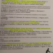
Poster P016: Octylisothiazolinone is a relevant nonoccupational contact allergen in leather goods and may show cross-reactivity to methyisothiazolinone.   J. Leysen et al.

Poster P020: Airborne bullous allergic contact dermatitis from MI contained in a glass shower screen cleaning spray.  M.A Pastor-Nieto et al.

Poster P021: Allergic contact dermatitis from nickel is prevented using a novel barrier cream.  Niklasson B and Isaksson M.  New nickel prevention cream!  “We present a male worker with ACD due to exposure to nickel-containing tools and where an active barrier cream containing a strong metal chelating agent helped solved the problem.   … The leather gloves were analyzed for the release of nickel ions: one glove released 0.4ug nickel cm2 and the other 0.2ug nickel cm2. … The barrier cream, NIK-L-BLOKTM (Chemotechnique), captures the nickel ions using a strong chelating agent, diethylenetriaminepentaacetic acid, in a special formulation that immobilizes the nickel ions (as well as cobalt and chromium ions), thereby preventing allergic contact dermatitis.”

Poster P022 The methyisothiazolinone epidemic: a pan- European prospective study JF Schewensen et al. “Patients were exposed to the following products containing MI (and could be exposed to more than one category): dish-washing liquids (n=32), shampoo (n=30), bath/shower gel (n=22).  … Thirteen experienced an allergic reaction in newly painted rooms: ACD [skin] (n=11), rhinitis [nose] (n=2) and conjuncitivitis [eye] (n=1).  Eight (4.7%) experienced reactions to other airborne exposures than paint, for example cleaning agents… EFFECTIVE REGULATION OF MI IN COSMETIC AND OCCUPATIONAL PRODUCTS IS NOT YET IN PLACE.  The current data demonstrate the URGENT need for PREVENTIVE actions.”

These poster abstracts are printed and the articles are forthcoming.  It is critical to work with manufacturers, consumers, patient advocates (medical providers) and legislators to protect!!!