Free article on Suspender Nickel dermatitis – prevention is the key

Review of: ‘ Suspender ’ Dermatitis and Nickel Sensitivity

Original article:  D. Calnan and G. C. Wells. (1956). Suspender Dermatitis and Nickel Sensitivity. British Medical Journal, 1(4978), p. 1265-1268.

Reviewed by Brittanya Limone, BS, MA, MSIII, Loma Linda University.

  • Historically, nickel allergy contact dermatitis was primarily associated with women working in industrial processes.
    • Calnan and Wells use a case of suspender dermatitis, one of the most common causes of nickel allergic contact dermatitis c. 1956, to highlight the prevalence of this condition amongst women regardless of their employment.
  • A dermatitis reaction is thought to occur after friction and sweat wear on nickel-containing products. These mediums gradually remove the nickel coatings and allow for nickel ion absorption across one’s skin.
    • In addition to suspenders, other everyday nickel-containing products that may induce an allergic contact dermatitis include watches, bra clasps, and earrings.
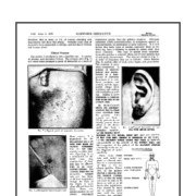
  • Typically, the first reaction site occurs in direct contact with the metal, also known as the primary site. This region appears as excoriated, superficial papules or a confluent patch.
    • Of note, pierced earrings were noted to cause earlobe dermatitis with crusts and exudates that might be mistaken for impetigo (infection).
  • Eruptions at sites distant to the metal’s direct contact are secondary sites. These occur in a symmetric fashion on the eyelids, sides of the neck, inner thighs, and elbow flexures.
    • Secondary reactions develop as papules or vesicles overlying an erythematous, edematous background with or without crusts and exudates.
  • A secondary flare-up is a more important clinical feature for diagnosis and treatment.
    • In terms of diagnosis, patients might not typically seek medical care until a secondary eruption. Therefore, recognition of these lesions, more commonly, leads to the diagnosis of nickel sensitivity.
    • However, conditions with secondary flare-up reactions are more difficult to treat. Patients with a primary lesion respond quickly to therapy, but once a secondary eruption occurs, clearing the condition is difficult and recurrences are more common.
  • Patch testing is used to confirm the diagnosis of nickel allergic contact dermatitis. However, waiting for the alleviation of an acute exacerbation is important as false positives from local reactions to patch testing may occur.


  • Prevention is key to this condition’s treatment and reduces recurrences.
    • The first step is the removal of all jewelry, metal clips or fasteners.
    • If nickel-containing products must be worn, then they should be covered with a protective coating of fabric, plastic, or enamel.
    • Alternatively, replacement products may be used such as items made of 100% plastic or nylon.

https://www.ncbi.nlm.nih.gov/pmc/articles/PMC1979680/

Nickel sensitivity and atopy

Early insight into the contributory role genetic factors play in the development of contact dermatitis

Synopsis of Nickel Sensitivity and Atopy (1964)

Original article:

A. Caron. (1964). Nickel Sensitivity and Atopy. British Journal of Dermatology, vol. 76: p. 384 – 387.

Reviewed by Brittanya Limone, BS, MA. MSIII, Loma Linda University.

  • “Atopy” is a genetic predisposition toward developing one or more of the following conditions: asthma, hay fever, urticaria, infantile eczema, or atopic dermatitis.
  • Determining the incidence of atopy has proven difficult because of reliance on clinical judgment and variable utilization of set parameters in making the diagnosis. For instance, the presentation of nasal congestion and recurrent sneezing episodes may be diagnosed as either hay fever, allergic rhinitis, or vasomotor rhinitis.
    • Several authors c.1964 estimated the prevalence of atopy in the United States’ general population being between 10-20%.
  • In the Caron case series, 37 patients with an established diagnosis of nickel contact dermatitis underwent further evaluation regarding their personal and family history of atopy, the results included the following:
    • Twenty-one (54%) had no history of atopy
    • Seven (19%) only had a family history.
    • Four (11%) had both a personal and family history
    • Five (14%) only had a personal history
  • The results from Caron’s case series suggested that the incidence of atopy amongst patients with nickel allergic contact sensitivity was no greater than its occurrence in the general population.
    • The frequency of atopy in 19% of family members was noted to be higher than expected for the general population but was attributed to the contributory role genetic factors play in the development of contact dermatitis.
  • This study concluded that nickel sensitivity occurs independently of atopy.

https://www.ncbi.nlm.nih.gov/pubmed/14201189

Only MCI/MI caused remarkable changes… skin cells affected

“only MCI/MI reduced NMF levels significantly… only MCI/MI caused remarkable changes at the microscopic level” to corneocytes (resident skin cells)…The altered corneocyte morphology suggests that skin barrier damage plays a role in the pathogenesis of MCI/MI contact allergy.”

IMPORTANT WORK!

Koppes SA1,2, Ljubojević Hadžavdić S3, Jakasa I4, Franceschi N5, Riethmüller C6, Jurakić Tončic R3, Marinovic B3, Raj N7, Rawlings AV7, Voegeli R8, Lane ME7, Haftek M9, Frings-Dresen MH1, Rustemeyer T2, Kezic S1.  Effect of allergens and irritants on levels of natural moisturizing factor and corneocyte morphology.  Contact Dermatitis. 2017 Mar 14. doi: 10.1111/cod.12770.

“Abstract
BACKGROUND:
The irritant sodium lauryl sulfate (SLS) is known to cause a decrease in the stratum corneum level of natural moisturizing factor (NMF), which in itself is associated with changes in corneocyte surface topography.
OBJECTIVE:
To explore this phenomenon in allergic contact dermatitis.

METHODS:
Patch testing was performed on patients with previously positive patch test reactions to potassium dichromate (Cr), nickel sulfate (Ni), methylchloroisothiazolinone (MCI)/methylisothiazolinone (MI), or p-phenylenediamine. Moreover, a control (pet.) patch and an irritant (SLS) patch were applied. After 3 days, the stratum corneum from tested sites was collected, and NMF levels and corneocyte morphology, expressed as the amount of circular nanosize objects, quantified according to the Dermal Texture Index (DTI), were determined.

RESULTS:
Among allergens, only MCI/MI reduced NMF levels significantly, as did SLS. Furthermore, only MCI/MI caused remarkable changes at the microscopic level; the corneocytes were hexagonal-shaped with pronounced cell borders and a smoother surface. The DTI was increased after SLS exposure but not after allergen exposure.

CONCLUSIONS:
MCI/MI significantly decreased NMF levels, similarly to SLS. The altered corneocyte morphology suggests that skin barrier damage plays a role in the pathogenesis of MCI/MI contact allergy.  DTI seems to differentiate reactions to SLS from those to the allergens tested, as SLS was the only agent that caused a DTI increase.”

https://www.ncbi.nlm.nih.gov/pubmed/28295421
© 2017 John Wiley & Sons A/S. Published by John Wiley & Sons Ltd

Nickel Earlobe Dermatitis – persistent sensitivity!

Nickel Earlobe Dermatitis

Original article:

Thomas L. Watt and Robert R. Baumann. (1968). Nickel Earlobe Dermatitis. Archives of Dermatology, vol. 98: p. 155-158.

Reviewed by Brittanya Limone, BS, MA. MSIII, Loma Linda University.

  • Although nickel is not a high potency sensitizer, increased frequency of nickel exposure, especially amongst young women, make it a common cause of allergic contact dermatitis.
  • The development of nickel sensitization can result in persistent sensitivity despite avoidance of items containing the metal. In addition, some patients may later have negative nickel patch testing but continue to have a dermatitis that stemmed from the nickel allergy.
  • Following repeated exposures, early nickel intolerances can eventually lead to metal rejection across the skin’s surface. This may lead to secondary site dermatitis reactions or a generalized dermatitis in some patients.
  • Watt and Baumann conducted a year-long observational study in 17 young women after they developed a draining ear lobe dermatitis 2-4 weeks after having ear lobe piercings with implanted earrings.
    • All of the women developed a positive eczematous reaction to nickel patch testing. Eleven had a personal history of atopy and three carried a family history.
    • Previously, the patients had tolerated nickel exposures to costume jewelry and clothing fasteners without trouble. The nickel allergy was only apparent following the earlobe piercing.
    • “In our experience, nickel sensitization following earlobe piercings is commonly mistaken for chronic infection, even though a similar dermatitis of the nonpierced earlobe is considered to be a cardinal sign of nickel allergy”.
  • The study found that despite the common perception that nickel dermatitis occurs more frequently after wearing “inexpensive” jewelry, nickel dermatitis was just as readily observed in these women following exposure to 14-karat gold and sterling silver earrings as with the inexpensively plated jewelry.
  • The study concluded that nickel earlobe dermatitis was highly associated with a personal history of atopy. Therefore, determination of a prior history of nickel allergy and atopy must be conducted prior to earlobe piercings.
    • Of note, careful screening for “latent atopics” must also be performed as they are equally susceptible to nickel earlobe dermatitis. These individuals may never have an obvious atopic disease but have a positive family history, positive skin testing, physical signs suggesting atopy, and bear atopic offspring.
  • According to Watt and Baumann, a history of nickel allergy is an “absolute contraindication” to earlobe piercings. On the other hand, personal history of atopic disease only requires warning patients about the possibility of nickel sensitization.

https://www.ncbi.nlm.nih.gov/pubmed/5667228

Role of low Nickel Diet in Nickel Allergy

What Role Does the low nickel diet Play in the Management of Nickel Allergy?

Original article:

Eleese Cunningham. What Role Does Diet Play in the Management of Nickel Allergy? Journal of the Academy of Nutrition and Dietetics, 2017; Vol. 117: p. 500.

Reviewed by Brittanya Limone, BS, MA. MSIII, Loma Linda University

  • Nickel is a common cause of allergic contact dermatitis (ACD) in the United States with rising detected prevalence. ACD reactions typically involve an ‘itchy rash’ following contact with nickel-containing compounds and are able to be confirmed using the diagnostic patch test.
  • Patients may present with widespread dermatitis, in the absence of contact with known allergens, raising the suspicion for dietary sources of nickel.
  • Notably, nickel amounts differ amongst plant types and also vary based on the levels of nickel present in the soil during growth. In the case of seafood, the water nickel content is a major contributing factor.
  • Eleese Cunningham reported that “In most studies, the richest sources of dietary nickel are found in nuts, dried peas and beans, whole grains, and chocolate.” Of note, the use of stainless-steel cookware and metal machinery may increase the nickel content in home-cooked meals and processed foods, respectively.
  • The pervasiveness of nickel across food groups makes a ‘nickel-free’ avoidance diet challenging. A diet high in iron-rich foods may be beneficial for patients with nickel sensitivities:
    • Iron and nickel compete for the same transport pathway during intestinal absorption. Consequently, if more iron is shuttled across intestinal mucosa, then less nickel will be transported.
    • In contrast, while normally only a small percentage of nickel is absorbed, this amount increases in patients with iron deficiencies. Additional processes that may lead to increased absorption of nickel include lactation or a disruption of the gastrointestinal barrier, such as in irritable bowel syndrome.1
  • Evidence suggests that nickel sensitive patients may benefit from reducing high nickel-containing foods from their diet.   Eleese Cunningham notes that the low-nickel diet should be followed for 4-6 weeks and then evaluate for improvement patient symptoms.

Additional Reference:

  1. Rizzi A, Nucera E, Laterza L, et al. Irritable Bowel Syndrome and Nickel Allergy: What Is the Role of the Low Nickel Diet? Journal of Neurogastroenterology and Motility. 2017;23(1):101-108.