Petrolatum Facts—150 years to today.

Petrolatum was discovered in 1859 inadvertently by the oil rig drillers in Titusville, Pennsylvania. The thick “rod wax” caused the rigs to malfunction and had to scraped off, however the workers found it to help soothe and heal their skin cuts. Robert Chesebrough, a chemist, refined, distilled, patented, and named it Vaseline—from the German wasser (water) and the Greek elaion (olive oil).

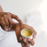
Purified petrolatum is a highly useful moisturizer and skin protectant as it prevents up to 98% of transepidermal water loss, is triple-purified and devoid of any allergens. It is recommended by the National Eczema Association as safe treatment option. There have not been any evidence-based reports directly linking purified petrolatum to cancer.

However, unrefined, low grade petrolatum does contain carcinogens called polycyclic aromatic hydrocarbons. Although the US does not regulate petrolatum use in cosmetics, labels stating “white petrolatum” or “Petrolatum, USP” can be sure they are buying a purified high grade product.

Petrolatum itself has never been reported to cause allergic contact dermatitis. Some petrolatum-based products, however, may not use a purified form of petroleum and often have additional ingredients such as Lanolin or fragrances and should be carefully evaluated for their ingredients by patients with known sensitizations.  

Free Article: IRRITABLE BOWEL SYNDROME-LIKE DISORDERS IN ENDOMETRIOSIS: PREVALENCE OF NICKEL SENSITIVITY AND EFFECTS OF A LOW-NICKEL DIET. AN OPEN-LABEL PILOT STUDY.

Authors: Borghini RPorpora MGCasale RMarino MPalmieri EGreco NDonato GPicarelli A

Journal: Nutrients. 2020 Jan 28;12(2). pii: E341. doi: 10.3390/nu12020341.

Abstract

Alimentary nickel (Ni) may result in allergic contact mucositis (ACM), whose prevalence is >30% and may present with IBS-like and extra-intestinal symptoms. These symptoms are also frequent in endometriosis, and Ni allergic contact dermatitis has already been observed in endometriosis. Therefore, intestinal and extra-intestinal symptoms in endometriosis may depend on a Ni ACM, and a low-Ni diet could improve symptoms. We studied the prevalence of Ni ACM in endometriosis and focused on the effects of a low-Ni diet on gastrointestinal, extra-intestinal, and gynecological symptoms. We recruited 84 women with endometriosis, symptomatic for gastrointestinal disorders. Thirty-one out of 84 patients completed the study. They underwent Ni oral mucosa patch test (omPT), questionnaire for intestinal/extra-intestinal/gynecological symptoms, and a low-Ni diet. Clinical evaluation was performed at baseline (T0) and after three months (T1). Twenty-eight out 31 (90.3%) patients showed Ni omPT positive results, with Ni ACM diagnosis, whereas three out of 31 (9.7%) patients showed negative Ni omPT. After three months of low-Ni diet, all gastrointestinal, extra-intestinal and gynecological symptoms showed a statistically significant reduction. Ni ACM has a high prevalence in endometriosis and a low-Ni diet may be recommended in this condition to reduce gastrointestinal, extra-intestinal and gynecological symptoms.

Link to article: https://www.ncbi.nlm.nih.gov/pubmed/32012984

Nickel allergy is associated with wheezing and asthma in a cohort of young German adults: results from the SOLAR study.

Article from University Hospital Munich – elucidating the details of nickel allergy by – Kolberg L1,2,3, Forster F1,3, Gerlich J1,3, Weinmayr G4, Genuneit J4,5, Windstetter D1, Vogelberg C6, von Mutius E3,7, Nowak D1,3, Drexler H8, Schäfer T9, Radon K1,3,10. ERJ Open Res. 2020 Feb 3;6(1). pii: 00178-2019. doi: 10.1183/23120541.00178-2019. eCollection 2020 Jan.

BACKGROUND: Nickel allergy is the most prevalent contact allergy. It belongs to a different hypersensitivity type to asthma and rhinoconjunctivitis. The aim of this analysis was to assess whether self-reported nickel allergy is associated with incident wheezing, asthma and rhinoconjunctivitis in young German adults, taking into account potential effect modification by sex.

METHODS: In total, 2051 (70.6%) participants aged 19-24 years took part in the second phase of SOLAR (Study on Occupational Allergy Risks), a follow-up study of ISAAC II (the second phase of the International Study of Asthma and Allergies in Childhood) in Germany. Self-reported nickel allergy, as well as having pierced ears, and the three outcomes incident wheezing, asthma and rhinoconjunctivitis, were analysed stratified for sex. Logistic regression adjusted for potential confounders was performed.

RESULTS: An association between self-reported nickel allergy and incident wheezing was observed for men and women, while only in males did pierced ears show a significant association with the outcome (adjusted OR 2.26, 95% CI 1.10-4.62). Also only in males, self-reported nickel allergy was associated with elevated odds for incident asthma (adjusted OR 4.34, 95% CI 1.22-15.41). Neither in men nor in women was a significant association observed for incident rhinoconjunctivitis.

CONCLUSION: Our results suggest that self-reported nickel allergy is associated with incident wheezing. Whether this association is due to environmental or genetic predisposition, or due to an overlap of the mechanisms of type I and type IV hypersensitivity, needs to be elucidated. https://www.ncbi.nlm.nih.gov/pubmed/32039258

Nickel Navigator App

Achieve success with your low nickel diet

In addition to the resources you can find at the Dermatitis Academy low nickel diet page, you can now increase your chances for success with the Nickel Navigator App. The app helps track your daily diet nickel intake! According to Rebelytics, the creators of the app, it is a “…support tool for individuals on a low-nickel diet.” The app helps you create a food journal to track your nickel intake. You can also browse though the food groups to see which foods are the lowest or highest in nickel, and you can search for a particular food and drill down into its data to understand how its nickel content varies. Rebelytics compiled this data from “more than 100 scientific sources, including national food studies and journal articles”. Interestingly, the average values can be weighted to the individual’s location so that the most geographically relevant sources are given priority. The app is easy to use, however for those that want to analyze the raw data this is readily available on their website.

TSW- awareness – one day at a time – TWO Free articles.

Thank you to the authors and the two great publishers for allowing this limited time free access.

“More research is required into long-term topical corticosteroid use and its discontinuation, including topical steroid withdrawal, particularly in the pediatric population”… writes Dr. Belinda Sheary (Australia) who recently published an article describing “[a] qualitative case series [that] studied 10 children whose parents stopped their chronic topical corticosteroid use and subsequently developed features typically reported in adults experiencing topical steroid withdrawal.”

https://www.medicaljournals.se/acta/content/abstract/10.2340/00015555-3144

And…

Also this, month the Journal of the Dermatology Nurses’ Association featured a case of Topical Steroid Withdrawal in their regular column: Teledermatology Viewpoint. “Although the exact mechanism of TSW has not yet been elucidated, some experts suggest that prolonged application of topical corticosteroids interferes with the production of nitric oxide, a potent vasodilator, in the endothelium (Rapaport & Lebwohl, 2003) … On the other hand, an alternative explanation of symptoms of TSW is that prolonged topical steroid application may inhibit cortisol production by keratinocytes, which may weaken the barrier function of the skin as well as result in hypersensitivity (Fukaya, 2016).”

https://journals.lww.com/jdnaonline/Fulltext/2019/03000/Burning_Red_Face.8.aspx

Moisturizer may enhance hydrocortisone absorption

Applying a moisturizing cream containing propylene glycol after applying topical hydrocortisone to the skin of patients with atopic dermatitis was shown to increase absorption of the glucocorticoid.

Original article:  Turpeinen,M.   Absorption of hydrocortisone from the skin reservoir in atopic dermatitis. British Journal of Dermatology. 1991; 124:358-360.

Reviewed by Allene Fonseca, PGY-3 Internal Medicine Resident Loma Linda University.

  • Turpeinen reported on the absorption rates of topical hydrocortisone applied to the skin of 7 patients (aged 4 months to 39 years) who were hospitalized for atopic dermatitis (AD) exacerbation.
    • Topical glucocorticoids were discontinued in all patients 36 hours prior to taking the absorption test.
    • A dexamethasone test was used to suppress morning cortisol secretion.
      • Dexamethasone is a strong steroid, 30-40x more potent than cortisol.
      • Administration of dexamethasone provokes (the body to have) a counter response of making less cortisol, because the homeostasis response sensed adequate corticoid in the system.
    • The serum level of cortisol of the 7 patients was checked after dexamethasone was given to suppress their natural cortisol production.
    • The dexamethasone suppression test was successful because all of the patients had below normal levels of plasma cortisol in the morning, compared to pre-treatment levels which were within normal limits.
      • To provide adequate suppression, the dexamethasone dose was adjusted to compensate for the percentage body surface area that had been receiving topical glucocorticoids previously.
        • For example, 0.25mg of dexamethasone was given for a body surface area of <0.5meters squared, and 2mg was given for a body surface area of >1.5 meters squared.
      • There were no noted side-effects of dexamethasone.
      • Subsequently, glucocorticoid absorption was determined by measuring serum cortisol levels, at set time intervals, beginning in the morning at 8:00-9:00, and subsequently 2h, 4h, 12h, 14h, 16h, and 24 h after the application of 1% hydrocortisone cream. In addition, the 3 adults’ serum was also checked again at 26 and 28 hours.
        • The cream was applied to the exact same areas that the topical hydrocortisone was applied too.
        • Children used 33-59g of topical hydrocortisone, and adults used 50-75g.
      • A moisturizing cream containing 80% water and 5% propylene glycol was applied to the hydrocortisone application sites, following the 12 hour blood sample.
      • In the adults, the moisturizing cream was again applied after the 24 hour blood sample was collected.
        • The changes in cortisol level between the different time intervals were statistically significant.
        • After applying 1% hydrocortisone once, plasma cortisol rose within 4 hours, and then steadily lowered.
      • In the 8 month old and the 3, 7 and 39 year old patients there was a rise in cortisol levels just 4 hours after applying the moisturizing cream, suggesting that within 4 hours the moisturizing cream can support increased absorption of topical steroid from skin reservoirs in some individuals.
      • In the 4 month old and the 19 and 20 year old patients there was not a significant rise in cortisol levels after applying the moisturizing cream
      • Following the 24 hour blood draw, the 3 adults again applied moisturizer.  There was no significant rise in serum cortisol levels. This finding suggests that applying moisturizer 24 hours after applying topical steroids does not increase the absorption of steroids.
    • This study suggests that topical hydrocortisone may be retained in the skin of patients with AD for up to 12 hours, and the absorption from the skin may be further enhanced by applying a moisturizing cream containing water and propylene glycol (PG).
      • PG, may work synergistically to increase the absorption of hydrocortisone (1).

References

  1. Turpeinen, M.   Absorption of hydrocortisone from the skin reservoir in atopic dermatitis. British Journal of Dermatology. 1991; 124:358-360.
  2. Zesch A. Schaeffer H. Penetration of radioactive hydrocortisone in human skin from various ointment bases. II. In vivo experiments. Arch Dermatol Forsch1975: 252: 245-56.


SNAS and thyroid autoimmunity risk

SNAS (Systemic nickel allergic syndrome may predispose susceptible individuals to developing chronic autoimmune thyroiditis (CAT)


Systemic nickel allergic syndrome as an immune-mediated disease with an increased risk for thyroid autoimmunity

Original Article:

Andrioli M, Trimboli P, Maio D, Persani L, Minelli M. Systemic nickel allergic syndrome as an immune-mediated disease with an increased risk for thyroid autoimmunity. Endocrine. 2015;50(3):807-810.

Go To Pubmed

Reviewed by Erin Wesner, BS, MSII & Jack Guccione, BS, MSIII. Loma Linda University School of Medicine


Keywords: Systemic nickel allergic syndrome, Nickel, Chronic autoimmune thyroiditis, Hashimoto


Key points:

  • Systemic nickel allergic syndrome (SNAS) may predispose susceptible individuals to developing chronic autoimmune thyroiditis (CAT)
  • Thyroid dysregulation is identified by elevated blood levels of thyroglobulin antibody and symptoms including fatigue, constipation, and weight gain.
  • Prior nickel sensitization with gastrointestinal and diffuse cutaneous symptoms and elevated thyroglobulin antibody may indicate SNAS-related thyroid disorder.
  • In over 200 patients studied, SNAS positive individuals showed a two-fold greater prevalence of CAT.


Introduction:

  • Chronic autoimmune thyroiditis (CAT) is the most common cause of hypothyroidism in areas with adequate iodine intake.1
    • CAT results from high circulating levels of anti-thyroid antigens and inflammatory mediators that cause gradual thyroid destruction.
    • Genetic, autoimmune and environmental factors (viruses, stress, hormones, pollution) have been identified as potential causes of CAT.2
  • Systemic nickel allergic syndrome (SNAS) is a chronic inflammatory disorder. It is characterized by a constellation of symptoms which can involve cutaneous, respiratory, gastrointestinal, and/or neurologic following systemic exposure to nickel (eg:  ingestion, implantation, inhalation) in individuals with prior nickel sensitization. (Table 1).1,3
    • This is in contrast to allergic contact dermatitis (ACD) wherein localized skin lesions erupt in response to direct contact with nickel (jewelry).
    • Approximately 20% of patients with a delayed allergy to nickel sulfate experience SNAS.1
    • SNAS is mediated through the release of cytokines by Th2 cells and Th1 cells, particularly IL-5 and IL-4.1,3
    • Intestinal biopsies taken from SNAS patients following nickel ingestion have shown increased amounts of CD4+ lymphocytes in the duodenal lamina propria and a decrease in the number of epithelial CD8+ lymphocytes due to apoptosis.3

Andrioli et al Article Review:

  • Andrioli et. al evaluated 239 patients over a 12-month period who had presented with immune-mediated inflammatory disease at the Department of Immune-Mediated Inflammatory Diseases of the Padre Pio Hospital in Campi Salentina, Lecce, Italy.1
    • All subjects underwent thyroid function testing as well as thyroid gland ultrasonography to identify thyroid disease.1
    • Thyroglobulin antibody (TGAb) (a blood test for thyroid dysfunction) was used to identify CAT in the study participants.
    • The authors defined SNAS by cutaneous and systemic manifestations, positive Ni patch testing in conjunction with an oral Ni challenge test, and clinical improvement following 4 weeks of a low Ni diet.1
      • 136 subjects met the diagnostic criteria for SNAS.1
      • Out of the remaining 103 subjects (non-SNAS), 40 tested positive for non-SNAS hypersensitivities (gluten, idiopathic dermatitis, lactose intolerance, allergic sensitivity to aeroantigens).1
      • The authors reported positive CAT in 26.5% of the SNAS group versus 12.7% in the non-SNAS group.
        • Notably, the global prevalence of CAT is estimated to be 1-2% in Caucasian women, but elevated in individuals with immune-mediated inflammatory disease, although the precise mechanisms remain unknown.1,4,5
        • For example, individuals with type 1 diabetes, celiac disease, Sjogren’s syndrome, rheumatoid arthritis and systemic lupus erythematosus all carry increased risk of developing CAT. None of these diseases were specifically identified in the non-SNAS group studied in Andrioli et al.1,6-9
      • Of note, TGAb levels were two fold higher in the SNAS group when compared to the non-SNAS group (19.9% vs. 7.8%).1


Key Points:

  • Andrioli et al. recognized a statically significant association between CAT and SNAS.1
  • The authors reported a two-fold greater prevalence of CAT in patients with SNAS (26.5%) compared to patients with other non-SNAS immune-mediated inflammatory disorders (12.7%).1
  • Based on Andrioli et al’s findings and literature review, the authors suggest that metal induced allergic reactions from nickel may promote increased production of TGAb through an autoimmune-inflammatory mechanism resulting in the development of CAT.
    • SNAS is associated with elevated levels of IL-4, a stimulator of B lymphocyte activity and differentiation.This cytokine may play a role in the production of TGAb in susceptible individuals when systemically exposed to nickel.
  • Concordant with these findings, elevated T-cell mediated pro-inflammatory cytokines are present in nickel-sensitized individuals, placing those with SNAS at a higher risk of developing autoimmune disorders (CAT).10
    • Further studies are warranted to define the pathogenic mechanism and protocol for screening for CAT in patients with SNAS.1


Table 1: Common SNAS and CAT individual and shared symptoms in descending order of frequency*10,11

Symptoms of SNAS10 Symptoms of CAT11 Shared symptoms10,11
Urticaria/angioedema Cold intolerance Fatigue
Dermatitis Impaired memory Edema
Recurrent abdominal pain Hair loss Constipation
Diarrhea Slowed movements Headache
Dyspepsia Brady cardia <60/min Bloating
Gastro-esophageal reflux Dry, coarse skin  
Itching Weight gain  
     
*SNAS – systemic nickel allergy syndrome; CAT – chronic autoimmune thyroiditis


References:

  1. Andrioli M, Trimboli P, Maio D, Persani L, Minelli M. Systemic nickel allergic syndrome as an immune-mediated disease with an increased risk for thyroid autoimmunity. Endocrine. 2015;50(3):807-810.
  2. Duntas LH. Environmental factors and thyroid autoimmunity. Ann Endocrinol (Paris). 2011;72(2):108-113.
  3. Braga M, Quecchia C, Perotta C, et al. Systemic nickel allergy syndrome: nosologic framework and usefulness of diet regimen for diagnosis. Int J Immunopathol Pharmacol. 2013;26(3):707-716.
  4. Weetman AP. Non-thyroid autoantibodies in autoimmune thyroid disease. Best Pract Res Clin Endocrinol Metab. 2005;19(1):17-32.
  5. Fallahi P, Ferrari SM, Ruffilli I, Elia G, Biricotti M, Vita R, Benvenga S, Antonelli A. The association of other autoimmune diseases in patients with autoimmune thyroiditis: Review of the literature and report of a large series of patients. Autoimmun Rev. 2016; 15(12):1125-1128.
  6. Appenzeller S, Pallone AT, Natalin RA, Costallat LT. Prevalence of thyroid dysfunction in systemic lupus erythematosus. J Clin Rheumatol. 2009;15(3):117-119.
  7. Biro E, Szekanecz Z, Czirjak L, et al. Association of systemic and thyroid autoimmune diseases. Clin Rheumatol. 2006;25(2):240-245.
  8. Counsell CE, Taha A, Ruddell WS. Coeliac disease and autoimmune thyroid disease. Gut. 1994;35(6):844-846.
  9. Piatkowska E, Szalecki M. Autoimmune thyroiditis in children and adolescents with type 1 diabetes. Pediatr Endocrinol Diabetes Metab. 2011;17(4):173-177.
  10. Ricciardi L, Carni A, Loschiavo G, et al. Systemic nickel allergy: oral desensitization and possible role of cytokines interleukins 2 and 10. Int J Immunopathol Pharmacol. 2013;26(1):251-257.
  11. Kostoglou-Athanassiou I, Ntalles K. Hypothyroidism – new aspects of an old disease. Hippokratia. 2010;14(2):82-87.


Delayed-type hypersensitivity to metals in connective tissue diseases

Delayed-type hypersensitivity to metals in connective tissue diseases and Fibromyalgia


Original article:

Bjørklund G, Dadar M, Aaseth J. Delayed-type hypersensitivity to metals in connective tissue diseases and fibromyalgia. Environmental Research. 2018;161:573-579. doi:10.1016/j.envres.2017.12.004

Reviewed by Jack Guccione, MS3 Loma Linda University School of Medicine.

Keywords: Fibromyalgia, Mixed connective tissue disease, Rheumatoid arthritis, Nickel, Mercury, Gold, Palladium, Metals, Allergy.

Abstract excerpt from: Delayed-type hypersensitivity to metals in connective tissue diseases

“Rheumatoid arthritis (RA) often involves the small joints of the hands in a symmetrical fashion that can lead to loss of joint function, and RA, as well as Sjögren’s syndrome (SS) and other rheumatic diseases, are often accompanied by sensitivity to metals… Metal-related DTH [delayed-type hypersensitivity] in these patients will induce an inflammatory response. Such inflammations are important factors in CTD [connective tissue disease] progress. It is hypothesized that metal-specific T cell reactivity can act as an etiological agent in the propagation and chronification of rheumatic inflammation. The key responses of metal delayed-type hypersensitivity in autoimmunity are precipitating as an appealing challenge for further investigations.”

Review:

  • Connective Tissue Disease (CTD) refers to a group of autoimmune diseases including rheumatoid arthritis (joint pain, malformation), Sjogren’s syndrome (excess tears and saliva), dermatomyositis, and systemic lupus erythematosus (joint pain, swelling).3
  • Fibromyalgia (FM) is a chronic rheumatic disease with an unknown cause that presents with tenderness and pain at 11-18 “trigger points” on the body and a constellation of symptoms that include cognitive and sleep impairment and widespread musculoskeletal pain and stiffness.
    • It is found in up to 6.6% in the general population, affecting women 2x more than men.1,2
    • There is significant overlap in non-musculoskeletal symptoms with chronic fatigue syndrome.
    • Persistent symptoms can lead to social inability, depression, and poor overall quality of life.1
  • Bjorklund et al. reviewed more than 100 studies and two case reports which had found positive associations between metal allergies (see table 1) and CTD or FM1
    • One study in the review reported THAT over 50% of women who tested positive for FM also self-reported having prior positive skin testing to nickel (bracelets, earrings).5
    • This is a significantly higher sensitization rate than is estimated in the US general population to nickel of 17-20%.6Similarly, two reviewed case reports anecdotally showed removal of nickel-containing dental appliances (bridges, wires) resulted in resolution of fatigue and FM pain in the patients.8
    • Bjorklund reviewed Hart et al.’s paper which found exposures to silica, mineral oils, and traffic pollution containing detectable levels of nickel, mercury, and palladium to be a significant risk factor for the development of RA.7
    • Bjorklund et al.’s review of a larger study (N=111), showed up to 76% of patients reported improvement of their chronic fatigue when dental metals containing nickel were removed.9
      • This study documented removal of mercury tooth-fillings resulted in substantial “health improvements” in individuals with FM including reduced migraine, less joint pain in wrists, hands, and fingers, and reduced fatigue symptoms.9
    • Another review was of an environmental study from 2007 on a community subdivision located near a petroleum tank battery.
      • The primary researchers found a significantly higher incidence of confirmed diagnosed SLE in the exposed community, compared to an unexposed near-by community.4
      • In this same study, ambient air testing found higher levels of petroleum hydrocarbons as well as increased levels of mercury in the air, which are known to suppress the immune system and lead to SLE.4
      • One of Bjorklund’s reviewed studies described the use of the ‘Memory Lymphocyte Immunostimulation Assay (MELISA) that detected delayed T-cell reactivity to dental-metals in patients suffering from FM.9
        • In this study, all patient with a history for FM also tested positive for at least one of the metals in their dental materials with MELISA
        • A 5-year follow up from removing these metals showed that half the patients no longer fulfilled the American college of rheumatology 1990 criteria for FM.9
        • An explanation for this finding is that sensitization to nickel or mercury in dental amalgams can cause higher levels of pro-inflammatory cytokines such as interferon-gamma when compared to healthy individuals.
        • Though not reviewed by Bjorklund, studies have found changes in free radical/antioxidant balance, as well as low levels of glutathione, correlate with metal induced inflammation, thus putting certain individuals at higher risk for developing FM.10
          • This remarkable finding suggests metal-induced inflammation may be reversible and is an important risk factor in patients with FM.
        • Bjorklund also reviewed literature regarding the potential protective effects of micronutrients in apparently alleviating autoimmune diseases..
          • For example, selenium was reported to potentially inhibits the toxic effects of exposure to other metals.11
          • These authors proposed low selenium in our diet can result in higher levels of other metals (mercury) thereby aggravating muscle pains seen in FM.12

Conclusion:

  • The authors review compelling findings from a variety of published studies that show patients suffering from CTD or FM may benefit from reduced exposure to certain metals.
    • These benefits include reduced inflammation and potential partial reversal of symptoms, which can ultimately help restore the efficacy of conventional therapies.


Table 1: Summary of metals and their relation to CTD and FM compiled from various papers reviewed in Bjorklund et al.

Metals and their relation to CTD and FM 1 What is it used for? What conditions or connective tissue disease is associated with it? What are the symptoms?
Gold 13 -Injectable Treatment for Rheumatoid arthritis14


-Jewelry, dental alloys, and orthopedic appliances 15


-chinaware16

-Gold allergy seen in 50% of RA patients studied (n=399) after treatment with gold salts14 Eczematous skin reaction 15
Palladium 15 -Dental Crowns, toothbrushing may increase palladium release, 17 Jewelry -Cross-reaction with nickel resulting in dermatitis 18 -Oral diseases such as lichen planus or stomatitis 19
Titanium 20 -Dental implants20 -Chronic Fatigue Syndrome, Multiple chemical sensitivity20 -Joint pain, nerve pain, chronic fatigue syndrome, depression, acne-like inflammation 20
Chromium 21 -Dye, detergent, cosmetics, leather, wood preservative 21

-Cement production 15

-Shoe contact dermatitis 22-Insulin dysregulation 23


-Bronchial asthma, hepatotoxicity 24


References:

  1. Bjorklund G, Dadar M, Aaseth J. Delayed-type hypersensitivity to metals in connective tissue diseases and fibromyalgia. Environ Res. 2018;161:573-579.
  2. Salaffi F, Sarzi-Puttini P, Girolimetti R, Atzeni F, Gasparini S, Grassi W. Health-related quality of life in fibromyalgia patients: a comparison with rheumatoid arthritis patients and the general population using the SF-36 health survey. Clin Exp Rheumatol. 2009;27(5 Suppl 56):S67-74.
  3. Iaccarino L, Gatto M, Bettio S, et al. Overlap connective tissue disease syndromes. Autoimmun Rev. 2013;12(3):363-373.
  4. Dahlgren J, Takhar H, Anderson-Mahoney P, Kotlerman J, Tarr J, Warshaw R. Cluster of systemic lupus erythematosus (SLE) associated with an oil field waste site: a cross sectional study. Environ Health. 2007;6:8.
  5. Regland B, Zachrisson O, Stejskal V, Gottfries CG. Nickel Allergy is Found in a Majority of Women with Chronic Fatigue Syndrome and Muscle Pain—and may be Triggered by Cigarette Smoke and Dietary Nickel Intake. Journal of Chronic Fatigue Syndrome. 2001;8(1):57-65.
  6. Thyssen JP, Menne T. Metal allergy–a review on exposures, penetration, genetics, prevalence, and clinical implications. Chem Res Toxicol. 2010;23(2):309-318.
  7. Hart JE, Laden F, Puett RC, Costenbader KH, Karlson EW. Exposure to traffic pollution and increased risk of rheumatoid arthritis. Environ Health Perspect. 2009;117(7):1065-1069.
  8. Muris J, Feilzer AJ. Micro analysis of metals in dental restorations as part of a diagnostic approach in metal allergies. Neuro Endocrinol Lett. 2006;27 Suppl 1:49-52.
  9. Stejskal V, Ockert K, Bjorklund G. Metal-induced inflammation triggers fibromyalgia in metal-allergic patients. Neuro Endocrinol Lett. 2013;34(6):559-565.
  10. De Luca C, Scordo MG, Cesareo E, et al. Biological definition of multiple chemical sensitivity from redox state and cytokine profiling and not from polymorphisms of xenobiotic-metabolizing enzymes. Toxicol Appl Pharmacol. 2010;248(3):285-292.
  11. Turner RJ, Finch JM. Selenium and the immune response. Proc Nutr Soc. 1991;50(2):275-285.
  12. Stoffaneller R, Morse NL. A review of dietary selenium intake and selenium status in Europe and the Middle East. Nutrients. 2015;7(3):1494-1537.
  13. Moller H. Dental gold alloys and contact allergy. Contact Dermatitis. 2002;47(2):63-66.
  14. Burmester GR, Blanco R, Charles-Schoeman C, et al. Tofacitinib (CP-690,550) in combination with methotrexate in patients with active rheumatoid arthritis with an inadequate response to tumour necrosis factor inhibitors: a randomised phase 3 trial. Lancet. 2013;381(9865):451-460.
  15. Forte G, Petrucci F, Bocca B. Metal allergens of growing significance: epidemiology, immunotoxicology, strategies for testing and prevention. Inflamm Allergy Drug Targets. 2008;7(3):145-162.
  16. Eisler R. Mammalian sensitivity to elemental gold (Au degrees). Biol Trace Elem Res. 2004;100(1):1-18.
  17. Wataha JC, Lockwood PE, Frazier KB, Khajotia SS. Effect of toothbrushing on elemental release from dental casting alloys. J Prosthodont. 1999;8(4):245-251.
  18. Faurschou A, Menne T, Johansen JD, Thyssen JP. Metal allergen of the 21st century–a review on exposure, epidemiology and clinical manifestations of palladium allergy. Contact Dermatitis. 2011;64(4):185-195.
  19. Durosaro O, el-Azhary RA. A 10-year retrospective study on palladium sensitivity. Dermatitis. 2009;20(4):208-213.
  20. Muller K, Valentine-Thon E. Hypersensitivity to titanium: clinical and laboratory evidence. Neuro Endocrinol Lett. 2006;27 Suppl 1:31-35.
  21. Thyssen JP, Strandesen M, Poulsen PB, Menne T, Johansen JD. Chromium in leather footwear – risk assessment of chromium allergy and dermatitis. Contact Dermatitis. 2012;66(5):279-285.
  22. Mortz CG, Andersen KE. Allergic contact dermatitis in children and adolescents. Contact Dermatitis. 1999;41(3):121-130.
  23. Ryan GJ, Wanko NS, Redman AR, Cook CB. Chromium as adjunctive treatment for type 2 diabetes. Ann Pharmacother. 2003;37(6):876-885.
  24. Shrivastava R, Upreti RK, Seth PK, Chaturvedi UC. Effects of chromium on the immune system. FEMS Immunol Med Microbiol. 2002;34(1):1-7.


FREE Access – Medical Necessity of Comprehensive Patch Testing

Free Access for 90 days, granted by publisher!!!

A Review of the Medical Necessity of Comprehensive Patch Testing

Zhu, Tian, Hao*; Suresh, Raagini; Warshaw, Erin‡§; Scheinman, Pamela; Mowad, Christen; Botto, Nina**; Brod, Bruce††; Taylor, James, S.‡‡; Atwater, Amber, Reck§§; Watsky, Kalman∥∥; Schalock, Peter, C.¶¶; Machler, Brian, C.***; Helms, Stephen†††; Jacob, Sharon, E.‡‡‡; Murase, Jenny, E.**§§§

Abstract:
Allergic contact dermatitis is associated with significant disease and economic burden in the United States. To properly manage allergic contact dermatitis, it is important to accurately identify the substance(s) implicated in the dermatitis to prevent disease recurrence. The commercially available T.R.U.E Test (36 allergens) screening panel has been reported to have a conservative hypothetical allergen detection rate of 66.0%, at most. Importantly, these calculations are based on the 78% of patients who had clinically relevant reactions to allergens present on the North American Contact Dermatitis Group screening series (70 allergens), without the use of supplemental allergens. Testing with supplemental allergens beyond a screening series can more fully evaluate an individual’s environmental and occupational exposure, which may significantly increase diagnostic accuracy. Comprehensive patch testing with additional allergens in sunscreens, cosmetics, and fragrances, for example, may increase the diagnostic yield as well as the likelihood of achieving a cure if the dermatitis is chronic and recalcitrant.
Free access provided by LWW for 90days.

Link to Access

Palladium allergic contact granuloma

Discussion on Palladium granuloma

Title:Palladium-induced allergic contact granuloma in a child wearing an earring

Original article:

Gonzalez-Perez, R, Carrillo-Ruiz, G, & Soloeta, R. Sarcoid-type Allergic Contact Granuloma Caused by Earrings in a Boy. Actas Dermo-Sifiliográficas (English Edition). 2012;103(1):73-74. doi: 10.1016/j.adengl.2010.12.008

Reviewed by Jack Guccione, BS. MSIII & Brittanya Limone, MA, BS, MSIV. Loma Linda University School of Medicine

Case Description

  • A case of granulomatous allergic contact dermatitis (ACD) in an 11-year-old boy due to an earring containing palladium reported in Spain.
    • He complained of a firm, asymptomatic papule on his left earlobe that had been present since he began wearing an earring 3-years prior.
    • Surgical removal and pathology evaluation of the papule revealed a reactive “sarcoidal granulomatous infiltration with no evidence of refringent material”, suggesting that the papule did not form due to retention of a foreign body (metal fragment) present in the skin.
    • Skin patch testing determined that the boy was sensitized to palladium.
  • Palladium chloride skin patch testing resulted in a primary eczematous response.
  • Mass spectrometry of the patient’s earring identified palladium as the primary metal composing the earring.
  • Given the temporal association between the skin patch testing and confirmation of palladium in the earring, the diagnosis of granulomatous allergic contact dermatitis reaction (gACDr) due to palladium was made [aka palladium granuloma].
  • The boy stopped wearing his earring and there was complete resolution of the reaction, confirming the diagnosis of palladium granuloma.


Discussion:

  • Sarcoidal-type (non-caseating/non-necrotizing) granulomas are abnormal nodules composed of inflammatory immune cells and macrophages.
    • They develop in response to a perceived need of the skin to encase a ‘foreign’ substance it is unable to eliminate.
    • This type of granuloma suggests the lesion is not caused by an infection from Mycobacteria tuberculosis, Treponema pallidum (syphilis) or some fungi, all of which can cause the necrotizing (internally breaking down) type of granuloma.
  • Several metals have been reportedly to cause gACDr (See Table 1).


Allergic contact granulomas are a less common presentation (variant) of allergic contact dermatitis.  The article was reviewed to acknowledge the spectrum of allergic dermatitis.


Table 1. Metals and Respective Sources Implicated in Granulomatous ACD

Metal Sources of Exposure
Palladium Earrings
Beryllium Mining

Fluorescent lighting tubes

Beryllium alloy production

Aluminum Vaccine & hypo-sensitization extract adjuvants

Deodorants

Tattoos

Zirconium Deodorants
Titanium Pacemakers
Mercury Tattoos
Chromium
Cobalt


References:

Gonzalez-Perez, R, Carrillo-Ruiz, G, & Soloeta, R. Sarcoid-type Allergic Contact Granuloma Caused by Earrings in a Boy. Actas Dermo-Sifiliográficas (English Edition). 2012;103(1):73-74. doi: 10.1016/j.adengl.2010.12.008

Lubeck, G., & Epstein, E. (1952). COMPLICATIONS OF TATTOOING. California Medicine, 76(2), 83–85.