It’s True – Metals in a Tattoo – Systemic Contact Dermatitis

Yes, indeed metals can be implanted in a tattoo… and systemically activated reactions can occur in those tattoos related to those metals…

“We recommend assessment of permanent tattoos for inflammation in all patients undergoing patch testing, for additional diagnostic correlation.”  [ 2008 Sep-Oct;19(5):E33-4]


Article 1 (came out yesterday) :de Cuyper C1Lodewick E2Schreiver I3Hesse B4Seim C5,6Castillo-Michel H4Laux P3Luch A3.   2017 Aug 9. doi: 10.1111/cod.12862. [Epub ahead of print]  Are metals involved in tattoo-related hypersensitivity reactions? A case report.

“BACKGROUND:  Allergic reactions to tattoos are not uncommon. However, identification of the culprit allergen(s) remains challenging.

OBJECTIVES: We present a patient with papulo-nodular infiltration of 20-year-old tattoos associated with systemic symptoms that disappeared within a week after surgical removal of metal osteosynthesis implants from his spine. We aimed to explore the causal relationship between the metal implants and the patient’s clinical presentation.

METHODS: Metal implants and a skin biopsy of a reactive tattoo were analysed for elemental contents by inductively coupled plasma mass spectrometry and synchrotron-based X-ray fluorescence (XRF) spectroscopy.

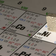
RESULTS: Nickel (Ni) and chromium (Cr) as well as high levels of titanium (Ti) and aluminium were detected in both the skin biopsy and the implants. XRF analyses identified Cr(III), with Cr(VI) being absent. Patch testing gave negative results for Ni and Cr. However, patch tests with an extract of the implants and metallic Ti on the tattooed skin evoked flare-up of the symptoms.

CONCLUSION: The patient’s hypersensitivity reaction and its spontaneous remission after removal of the implants indicate that Ti, possibly along with some of the other metals detected, could have played a major role in this particular case of tattoo-related allergy.”


Article 2: Cobalt tattoo reaction:

2017 Jun 1;15(3):221-222. eCollection 2017.  Chemical Tattoo Treatment Leading to Systemic Cobalt Hypersensitivity.  Zajdel NJ1, Smith WA2, Taintor AR3, Jacob SE4, Olasz EB5.

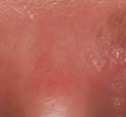
“An otherwise healthy 36-year-old Caucasian woman, without prior history of atopic dermatitis or eczema, presented to an outside dermatologist with a generalized, severely pruritic eruption involving the entire body except the face. One month previously, she had used a 50% trichloroacetic acid tattoo removal solution on a blue-colored tattoo on the medial aspect of the left ankle. The patient’s eruption persisted for 7 months, and after several attempts to slowly taper her prednisone dose, she presented to our institution. On physical examination, there was a 3-cm erythematous, lichenified plaque surrounding the tattoo (Figure). On the trunk and upper regions of the arms, there were scattered, 1- to 2-cm, nummular patches and plaques. Biopsy of a truncal lesion revealed spongiotic pustules with a mixed dermal infiltrate and scattered eosinophils, consistent with subacute spongiotic dermatitis.”


Article 3:  Systemic Dermatitis following surgery — presenting as tattoo reaction

2017 Jul 19;3(4):348-350. doi: 10.1016/j.jdcr.2017.05.003. eCollection 2017 Jul.  Systemic contact dermatitis to a surgical implant presenting as red decorative tattoo reaction.
“The patient reported that within 2 weeks of surgery, the red-containing areas of her tattoos, which were previously flat and uninflamed, became raised and pruritic.”…
Read this article
Article 4: SCD to chromate in a tattoo triggered by patch testing
2008 Sep-Oct;19(5):E33-4.Inflammation in green (chromium) tattoos during patch testing.  Jacob SE1, Castanedo-Tardan MP, Blyumin ML.

“We report three patients with permanent tattoos and chronic dermatitis. During patch testing, the patients’ dermatitis worsened, and the previously quiescent green-colored portions of the tattoos became inflamed. All three patients were patch-tested and had positive reactions to potassium dichromate 0.25% in petrolatum. Avoidance led to the resolution of both the dermatitis and the tattooinflammation. We recommend assessment of permanent tattoos for inflammation in all patients undergoing patch testing, for additional diagnostic correlation.”

Article 5: 1962
1962 Aug-Sep;74:288-94.Green tattoo reactions associated with cement dermatitis.
And … one of those reads that just makes you think… Article 6:
2004 Aug 21-27;364(9435):730.  A red tattoo and a swordfish supper.
Read more here
“Tsuruta et al. report- ed a case of a 40-year-old Japanese man with a red tattoo who developed a whole-body rash after eating 250 g of raw swordfish and alfonsino.”
Researchers are investigating metal allergic dermatitis and the role of piercing in nickel allergy.  Please pass along this survey.

Fiddler’s Neck and Nickel

Review of: Fiddler’s neck: Chin rest-associated irritant contact dermatitis and allergic contact dermatitis in a violin player

Original Article: Caero, Jennifer E & Cohen, Philip R. (2012). Fiddler’s neck: Chin rest-associated irritant contact dermatitis and allergic contact dermatitis in a violin player. Dermatology Online Journal, 18(9).

Reviewed by: Jacqueline Chen, BA. MSII

  • String players such as violinists and violists can develop dermatitis (inflammatory skin disease), colloquially called ‘Fiddler’s Neck, which may occur on the submandibular region (just below the jaw) or the supraclavicular (on the neck) region.
    • Submandibular Fiddler’s neck, or Fiddler’s neck type 1, refers to the irritation caused by mechanical frictional that occurs following prolonged contact with the chinrest.
      • Submandibular Fiddler’s neck is often described as a lichenified (thickened) plaque that may be darker than the surrounding skin.
      • Four main factors contribute to Fiddler’s neck Type I: pressure of the fiddle, friction, hygiene, and the instrument position.
      • A barrier cushion and adjustment to more horizontal positioning of the instrument has been recommended between the chinrest and submandibular neck to avoid fiddler’s neck type 1
    • Supraclavicular Fiddler’s neck, or Fiddler’s neck Type II occurs from contact allergy to the materials in the chinrest apparatus on the instrument.
      • Supraclavicular dermatitis usually presents as a pruritic (itchy) and erythematous (red) eczematous plaques.
      • Allergic contact dermatitis (the allergic skin response) to the chinrest is most often caused by nickel sulfate in the bracket that holds the chinrest to the instrument, but can also be caused by allergens in the composite woods.
      • The diagnosis of Fiddler’s neck Type 2 is often made based on the patient history and through observing the musician playing his/her instrument to evaluate the contact point of the rash with the instrument.
      • A patch test is the diagnostic test to confirm allergic contact dermatitis
      • Notably, sweat can dissolve nickel, contributing to its corrosion and increasing nickel absorption by skin.
      • To confirm nickel release, the dimethylglyoxime (DMG) test can be used
      • https://www.youtube.com/watch?v=dJFcHo5fDbY
      • Treatment of supraclavicular Fiddler’s neck should be focused on avoidance of the component in the chinrest containing the allergenic material.


To read the article by Caero and Cohen please click here.

If you suffer from an allergy to your musical instrument, and would like to participate in a nickel allergy awareness survey, please click here.

Nickel allergy – immunologic inflammatory pathways

Review of: Nickel sulfate promotes IL-17A producing CD4+ T-cells by an IL-23 dependent mechanism regulated by TLR4 and Jak-STAT pathways

Original article: Bechara, R, Antonios, D, Azouri, H, Pallardy, M, Nickel sulfate promotes IL-17A producing CD4+ T-cells by an IL-23 dependent mechanism regulated by TLR4 and Jak-STAT pathways. The Journal of Investigative Dermatology. 2017 Jun 17.

Reviewed by: Jacqueline Chen, BA. MSI & Brittanya A. Limone, MS, BS. MSIV

  • Allergic contact dermatitis (ACD) is classically described as a Type IV hypersensitivity reaction, however, the distinctive characteristics of a nickel-induced allergic contact dermatitis (Ni-ACD) lead to immunologic mechanisms that not only encompass a Th1 response but involve additional inflammatory cells, cytokines, and pathways.
  • In Ni-ACD, dendritic cells (antigen presenting [accessory] cells) play a critical role. Dendritic cells bind the antigenic nickel absorbed in the skin and then present it to T-cells at local lymph nodes, coordinating T-cell differentiation through cytokine messengers.
    • The two most crucial cytokine signals include:
      • IL-12p70 which promotes a T-helper Cell 1 type (Th1) response
      • IL-23 which stimulates the development of T-helper Cell 17 type (Th17) cells
    • Notably, the presence of IL-17A produced by Th17 cells correlates with the clinical reaction in nickel allergic patients. An injection of anti-IL-17 neutralizing antibodies may limit the severity of the contact hypersensitivity.
    • The IL-23/IL-12p70 balance determines the primary immunologic mechanism of the hypersensitivity reaction.
      • Increases in the IL-23/IL12p70 balance lead to a greater Th1 cell polarization
      • Decreases in the IL-23/IL-12p70 ratio produce a stronger Th17 cell response.
    • Brechara et al identified 5 specific modulators of T-cell differentiation that are important in the development of Ni-ACD through alterations in the IL-23/IL-12p70 balance.
      • IFN-γ
        • Produced by Th1 cells.
        • Greatly increases the IL-23 levels produced by nickel sulfate (NiSO4)-treated dendritic cells.
        • The increase in the IL-23/IL-12p70 ratio favors Th17 cell development.
      • Jak-STAT pathway
        • Inhibition of the Jak-STAT pathway increases IL-23.
        • Alternatively, activation of the pathway will increase IL-12p40 and IL-12p70 levels and decrease the IL-23/IL-12p70 balance.
        • This decrease in the IL-23/IL-12p70 balance favors a Th1 cell response.
      • TLR4, p38MAPK and NFkB pathways
        • Activation of these pathways is essential for nickel-induced production of IL-23, IL-12p40 and IL-12p70.
        • Since both IL-23 and IL-12 cytokines are produced, the IL-23/IL-12p70 balance remains high.
      • In summary, Ni-ACD is a complex immunologic disease involving not only a cell-mediated Th1 response but also Th17 cell development with alterations in IFN- γ levels and TLR4, Jak-STAT, p38MAPK, and NF-kβ immunologic pathways.

Article: link to publishers site

Researchers are investigating the role of piercings and the development of nickel allergy – please consider to take the Loma Linda University Nickel Allergy Survey:

Nickel allergy survey


airborne allergic contact dermatitis- isothiazolinones is not rare

2017 Apr 27. doi: 10.1111/cod.12795. [Epub ahead of print]

Airborne allergic contact dermatitis caused by isothiazolinones in water-based paints: a retrospective study of 44 cases.

Abstract

BACKGROUND:

Airborne allergic contact dermatitis caused by paints containing isothiazolinones has been recognized as a health hazard.

OBJECTIVES:

To collect epidemiological, clinical and patch test data on airborne allergic contact dermatitis caused by isothiazolinone-containing paints in France and Belgium.

METHODS:

A descriptive, retrospective study was initiated by the Dermatology and Allergy Group of the French Society of Dermatology, including methylchloroisothiazolinone (MCI)/methylisothiazolinone (MI)- and/or MI-sensitized patients who developed airborne allergic contact dermatitis following exposure to isothiazolinone-containing paint.

RESULTS:

Forty-four cases were identified, with mostly non-occupational exposure (79.5%). Of the patients, 22.5% of also had mucosal symptoms. In several cases, the dermatitis required systemic corticosteroids (27.3%), hospitalization (9.1%), and/or sick leave (20.5%). A median delay of 5.5 weeks was necessary to enable patients to enter a freshly painted room without a flare-up of their dermatitis. Approximately one-fifth of the patients knew that they were allergic to MI and/or MCI/MI before the exposure to paints occurred.

CONCLUSION:

Our series confirms that airborne allergic contact dermatitis caused by paints containing isothiazolinones is not rare, and may be severe and long-lasting. Better regulation of isothiazolinone concentrations in paints, and their adequate labelling, is urgently needed.

https://www.ncbi.nlm.nih.gov/pubmed/28449346

Patch Testing in Hand Eczema – Free Article!

REVIEW of: The Ecacy of the Patch Test in Diagnosing Hand Eczema

Original article: Vigneshkarthik, N, Ganguly, S, and Kuruvila, S. Patch Test as a Diagnostic Tool in Hand Eczema. Journal of Clinical Diagnostic and Research, 2016; 10(11): WC04–WC07. India

Reviewed by Jacqueline Chen, BA. MSI and Brittanya Limone, MA. MSIII, Loma Linda University.

  • Hand eczema is a frequently encountered dermatologic problem that may be secondary to an allergic contact dermatitis (ACD). Patch testing is a diagnostic tool that can confirm an ACD etiology and determine the necessary allergens to avoid to prevent future exacerbations.
  • Vigneshkarthik, Ganguly, and Krurvila’s cross-sectional study analyzed patch test results to determine common allergens associated with ACD in patients with hand eczema. The study included 54 individuals, 27 men and women. The authors discovered that:
    • Of the patients with hand eczema 37% had positive patch test results to 25 different allergens, confirming that ACD is a common cause of hand eczema
    • Specific occupations were more susceptible including housewives, agriculturists, students and masons
  • Nickel was the most common allergen associated with hand eczema, particularly amongst patients with a vesicular hand eczema, or pompholyx, morphology.
    • In patients with nickel allergy, high levels of nickel in the diet have been associated with this more severe form of hand eczema.
  • Risk factors thought to be associated with the development of nickel hand eczema:
    • Ear piercings and adornment with nickel jewelry.
    • Chores in wet environments
    • Use of nickel-containing utensils
  • Detergents were another important association with nickel allergy
    • Over one-third of patients with regular detergent exposure had a positive patch test result.
      • 54% of these positive patch test results were due to nickel, either alone or in combination with cobalt sensitivity.
      • The association of nickel and cobalt allergies was determined to be due to co-exposure to the metals from a common source.

PubMed Link:  https://www.ncbi.nlm.nih.gov/pmc/articles/PMC5198439/   Adobe PDF Icon

Free Article on Topical Steroid addiction – withdrawal

Free article on topical steroid addiction – “Cortisol production by keratinocytes [skin cells] might work to regulate or moderate the friction between the outer environment and inner immune system by suppressing excessive inflammation or immune reaction. However, prolonged or excessive use of TCS induces skin atrophy which can make barrier function weak. Moreover, the decreased self-production of cortisol by the keratinocytes can cause hypersensitivity. The author considers it is one of the mechanisms of TSA or rebound phenomenon after TSW.”

Fukaya M1. .Histological and Immunohistological Findings Using Anti-Cortisol Antibody in Atopic Dermatitis with Topical Steroid Addiction.   2016 Mar;6(1):39-46. doi: 10.1007/s13555-016-0096-7. Epub 2016 Feb 2.  Dermatol Ther (Heidelb)

“Abstract

INTRODUCTION:

Though topical steroid addiction (TSA) in patients with atopic dermatitis (AD) has been recently discussed as a clinical problem, there are very few studies about its mechanism. The purpose of this study was to elucidate histological and immunohistological characteristics of TSA using anti-cortisol antibody.

METHODS:

Skin biopsy specimen from eight patients with AD was stained by anti-cortisol antibody (Biorbyt, orb79379). Subjects consisted of a child patient with a short history of topical corticosteroids (TCS) application, an adult patient with a long history of TCS application, and six adult patients who have experienced topical steroid withdrawal (TSW) and the rebound phenomenon.

RESULTS:

The staining in the epidermis by anti-cortisol antibody presented patchy defects in the child patient, the patient with a long history of TCS application, and two patients at the rebound period. Parakeratosis with poor formation of corneal layer was obvious in the child patient, the patient with a long history of TCS application, two patients recovered from TSA, and two patients at the rebound period.

CONCLUSION:

Prolonged application of TCS might suppress the cortisol production of keratinocytes which is poorly developed at the early ages before childhood and completed naturally as to growth. Rebound phenomenon after TSW can occur due to the relative insufficiency of cortisol in the epidermis and the immature corneal layer formation.”

https://www.ncbi.nlm.nih.gov/pmc/articles/PMC4799038/

Not all nickel allergy reactions are delayed

Nickel allergy early reactions reported within 30 min of contact!

 Nickel allergy in a Danish population 25 years after the first nickel regulation.

Author information

 Contact Dermatitis. 2017 Apr 7. doi: 10.1111/cod.12782. [Epub ahead of print]

Abstract

BACKGROUND:

Nickel in metallic items has been regulated in Denmark since 1990; however, 10% of young Danish women are still sensitized to nickel. There is a need for continuous surveillance of the effect of regulation.

OBJECTIVES:

To identify current self-reported metallic exposures leading to dermatitis in nickel-allergic patients, and the minimum contact time needed for dermatitis to occur.

METHODS:

A questionnaire was sent to all patients who reacted positively to nickel sulfate 5% pet. within the last 5 years at the Department of Dermatology and Allergy, Gentofte Hospital.

RESULTS:

The response rate was 63.2%. Earrings were the foremost cause of dermatitis after the EU Nickel Directive had been implemented, followed by other jewellery, buttons on clothing, belt buckles, and wrist watches. Dermatitis reactions within 10 min of contact were reported by 21.4% of patients, and dermatitis reactions within 30 min of contact were reported by 30.7% of patients. [Noting nickel allergy early reactions]

CONCLUSIONS:

Nickel exposures that led to implementation of a nickel regulation seem to persist. The durations of contact with metallic items to fall under the current REACH regulation of nickel correspond well with the results of this study.

KEYWORDS:

EU directive; allergic nickel dermatitis; metallic items; nickel; prolonged direct contact

TSW

Facing topical steroid withdrawal TSW – health matters

TSW – topical withdrawal syndrome

Facing up to withdrawal from topical steroids 

By Mary C. Smith, RN, MSN; Susan Nedorost, MD; and Brandie Tackett, MD

“Topical corticosteroids applied to the face to treat these symptoms can cause steroid rosacea and steroid addiction syndrome, resulting in new symptoms that perpetuate the topical steroid usage.”  “withdrawal … which is called steroid addiction syndrome.”

” The best time to prevent … is when topical corticosteroids are first prescribed.”

” Getting the red out

” Uncovering steroid rosacea

” Stopping the cycle

” Patient teaching

Call to Action: “Learn to recognize this condition”

Get article here


Nickel in Cocoa – speciation

Food Chem. 2017 Sep 1;230:327-335. doi: 10.1016/j.foodchem.2017.03.050. Epub 2017 Mar 10.

Nickel speciation in cocoa infusions using monolithic chromatography – Post-column ID-ICP-MS and Q-TOF-MS.

Abstract

Nickel (Ni) is considered to be a potentially harmful element for humans. Its levels in foodstuffs are normally low (below 0.2mgkg-1), but sensitive individuals may develop allergy to Ni as a result of dietary consumption. Cocoa contains relatively high Ni concentrations (around 3mgkg-1). Ni bioavailability, its role in the flavour of food and its potential impact on human health depends primarily on its chemical species. However, there is a lack of information about Ni speciation in cocoa. In this work Ni species were separated on a weak convective interaction media diethylamine (CIM DEAE) monolithic chromatographic column and quantified by the post-column isotope dilution inductively coupled plasma mass spectrometry (ID-ICP-MS). The Ni binding ligands in the separated fractions were identified “off line” by quadrupole time-of-flight mass spectrometry (Q-TOF MS). Ni was found to be present in the cocoa infusions as Ni2+ and Ni-gluconate and Ni-citrate complexes.

Triggers of ACD – allergens and the jewelry addict

Discussion of Triggers of Allergic Contact Dermatitis in Accessories – the Jewelry addict

Original article: Nanette B. Silverberg. (2016). The “Jewelry Addict”: Allergic contact dermatitis from repetitive multiple children’s jewelry exposures. Pediatric Dermatology 33: e103-e105

Reviewed by Lauren A. Ivey, MS. MSI & Brittanya A. Limone, MA, BS. MSIII Loma Linda University

  • Nickel Allergic Contact Dermatitis (Ni-ACD) can be a distressing problem, especially for young girls with pierced ears and a love for costume jewelry.
  • Nickel is the most commonly confirmed contact allergen in both children and adults.
  • Confirmed Ni-ACD has increased 3 to 4-fold since 1986.
    • Common sources of nickel exposure in children include jewelry and adornments, electronics, and school chairs.
    • Girls are especially at risk to early nickel exposure through earrings and daily us of costume jewelry (aka “jewelry addict”)
    • Electronics, eg cell phones, laptops, and tablets have increased the frequency of nickel exposure.
  • Silverberg presents a case study of a 9-year-old girl “addicted to costume jewelry” who developed ACD after exposure to different metal- and rubber-containing accessories.
    • Confirmed sources of the allergen exposure included rubber bracelets, cheaper metal jewelry, lip balm case and belt buckles.
    • Physical examination: classic involvement of the antecubital fossa (crease of arms) consistent with a diagnosis of atopic dermatitis, and lichenified plaques on the fingertips and dorsal hands.
    • Notable plaques were seen on the lips and perioral region associated with application of lip balm kept in the metal case.
    • Patch testing revealed a 3+ reaction to nickel (papular variant), 2+ to gold thiosulfate, and 3+ to thiuram, all of which were deemed clinically relevant considering the child’s history with jewelry containing these allergens and her  presentation.
  • Parents and caregivers must remain cognizant of important allergens in jewelry.
  • Because virtually any type of jewelry can be a source of allergy, children who exhibit persistent dermatitis should be tested for suspected allergens based on history and exposure.